Microscopy and instrumental methods in biology
This course overviews modern measurements and imaging techniques used in biophysics, molecular and cell biology. Its main goal is to teach students about the capabilities and limitations of each of the most popular techniques, and explain how to select the simplest and the most effective approach to answer a particular experimental question. Practical works will include a complete workflow of processing real experimental datasets from fluorescence and CD experiments, planning chromatography experiments, advanced work with fluorescence microscopy images using ImageJ and Pyton
Registration is open and will be possible until April 23. (closed)
Light absorption. Color. Spectrophotometers. Absorption-based methods. Dipole moment of molecules and absorption wavelength. (Shvadchak V.).
Principles of fluorescence. Jablonsky diagram. Fluorescence quantum yield. Fluorophores. Tryptophan and other natural fluorophores. GFP. Brightness. Solvatochromism. (Shvadchak V.).
Protein labeling. Cys-, Lys- reactive dyes. Intercalating dyes. FRET and its application to study protein interactions. Polarized light. Fluorescence anisotropy. (Shvadchak V.).
CD spectroscopy to determine protein structure. IR spectroscopy. (Shvadchak V.).
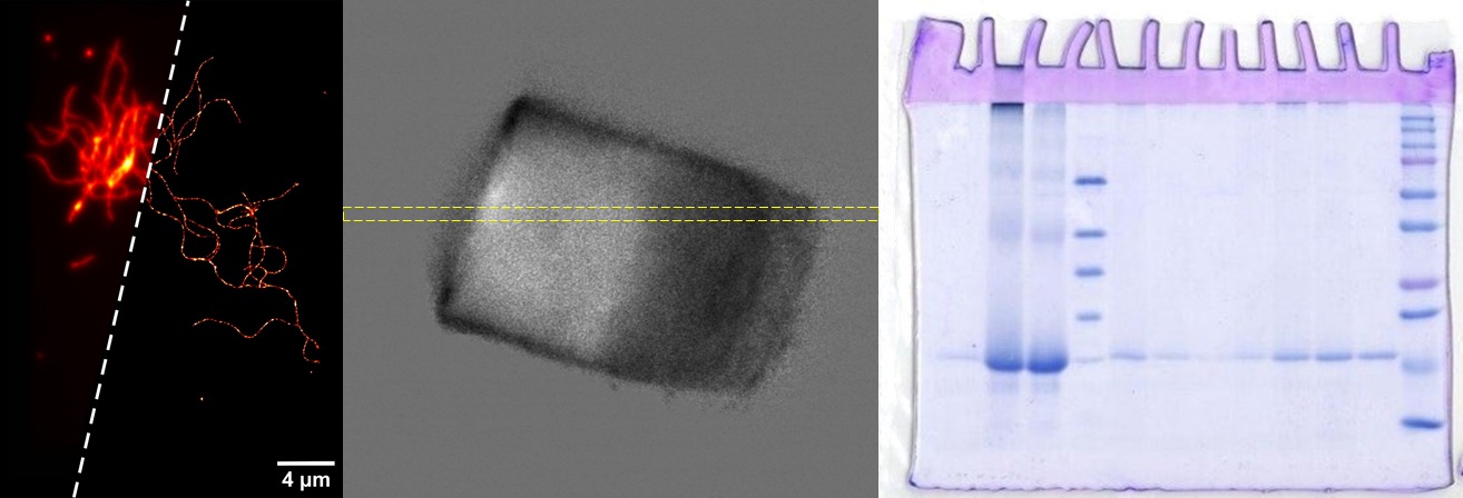
Electrophoresis of proteins and oligonucleotides. DLS. FCS. FCCS. (Shvadchak V.).
HPLC principle, preparative and analytical applications. Types of columns. Ion exchange chromatography. Size-exclusion. (Shvadchak V.).
Mass-spectrometry. LC-MS. ESI, MALDI and other ionization methods. Types of mass detectors. Fragmentation. LC-MS in proteomics. (Shvadchak V.).
Transmission microscopy, phase contrast. Fluorescence microscopy. Principal schemes of microscopes. Lasers. Filters. Dichroic mirrors. Channels. Digital image collection. Image resolution, micrones and pixels. Confocal microscopy. Z-slices. (Kovalchuk Yu.).
Membrane trackers, staining of nuclei. ImageJ/Fiji. Colocalization. TIRF. (Kovalchuk Yu.).
FRET and detection of interactions in microscopy. Fluorescence lifetime and FLIM. Diffraction limit. Superresulution, STORM. PALM. Application to image actin fibrils. (Shvadchak V.)
Fluorescent proteins. Small molecule dyes. Channel crosstalk and selection of fluorophores. Photodegradation during measurements. FRAP. Light intensity and damage to cells. Caged molecules and controllable photorelease. Photoswithchable molecules. (Shvadchak V.)
(Khoroshyy P.)
(Khoroshyy P.)
(Khoroshyy P.)
Principle. Resolution. Modes. Sample preparation. (Bondarenko N.)
(Bondarenko N.)
Principle and scheme of microscopes. XY and Z resolution. Sample preparation. Scanning speed and sample damage. Application for protein unfolding. (Shvadchak V.)
Spin. 13C and 15N protein labeling. NMR for protein structure analysis. Solid state NMR. ESR and free radicals. (Shvadchak V).
Protein crystallization. SAXS. (Shvadchak V.).
(Shvadchak V.)
Kd determination. Origin software for non-linear fitting.
(Shvadchak V.)
(Shvadchak V.)
(Shvadchak V.)
(Shvadchak V.)
(Shvadchak V.)
Fiji ImageJ - безкоштовна програма для обробки файлів даних з мікроскопів (завантаження Fiji).
Підручник по Image J
(Shvadchak V.)
(Khoroshyy P.)
(Bondarenko N.)
- Peter Jomo Walla Modern Biophysical Chemistry Detection and Analysis of Biomolecules, Wiley-VCH 2014 // розділи 1,2
- Пивоваренко В.Г. Абсорбційна та флуоресцентна спектроскопія органічних сполук; К.: ВПЦ «Київський університет», 2023
- Каталог спектрів поглинання та флуоресценції барвників
Сторінка завантаження програми VMD https://www.ks.uiuc.edu/Development/Download/download.cgi?PackageName=VMD
Cайт де можна скачати файли структур молекул з координатами атомів https://www.rcsb.org/ (в форматі PDB).