Histological Methods in Life Sciences
The course focuses on the contemporary histological methods in biomedical research. These include histology, immunohistochemistry/immunofluorescence, FISH, and in vivo staining techniques. The course teaches how to efficiently collect and process fresh-frozen and formalin-fixed specimens, and how to conduct paraffin- and resin-embedding.
Its objective is to provide or improve practical skills of students in histological techniques, and empower their relevant application in biological and medical research.
Why do we need histology? Development of histological techniques. Histological laboratory: general issues of location, organization and infrastructure. Equipment of the histological laboratory. Safety in the histological laboratory. Documentation and accounting. Features of procurement of equipment and reagents.
Methods for collecting and fixing samples (grossing). Decalcification of samples. Methods of histoprocessing. Infiltration and embedding. Histoprocessing of unfixed material (fresh-frozen sectioning). Microtomy.
General principles of histological staining techniques. Basic techniques of histological staining. Hematoxylin: basic formulations. Hematoxylin-eosin staining: a routine method in histological practice. Staining methods for bacteria and fungi. Connective tissue staining. Methods of nervous tissue staining. Enzyme histochemistry.
Bone tissue processing strategies. Histological processing of native (non-mineralised) bone. Grinding using the EXACT® system. Assessment of the bone-implant interface. Assessment of the implant surface. In vivo (vital) staining of the bone tissue. Decalcification.
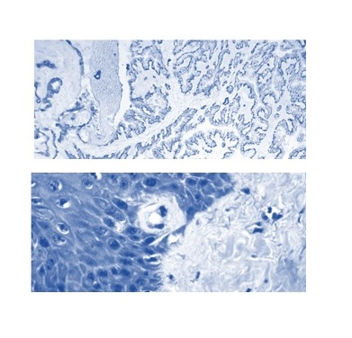
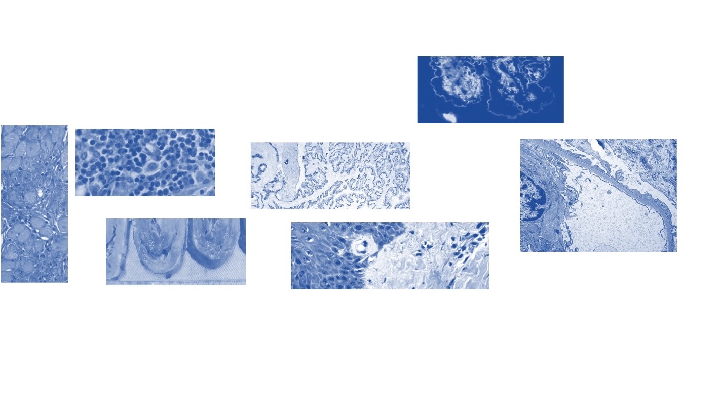
General principles of immunohistochemical method. Production of specific antibodies. Antigen retrieval. Blocking of endogenous peroxidase. Blocking of nonspecific antibody binding. Visualization. Immunofluorescence technique.
History of the DNA hybridization method. Principle of the in situ hybridization method. Opportunities and challenges. In situ hybridization in the diagnosis of malignant tumors. Detection of ERBB2 (HER2/neu) amplification as a powerful prognostic tool in the treatment of breast cancer. Prospects for use.
DNA extraction. Polymerase chain reaction. Sequencing. Data analysis methods.
The principle of operation of a light microscope. Modifications of the transmission light microscope. The video demonstrates the operation of the AXIO Imager A2 microscope in light-optical and fluorescence modes, and provides an example of image processing in the ZEN program.
I
https://forms.gle/
Регламент:
Вікно тестування: 9:00 - 16:00, 1 спроба, 30 хв.
Перебільшення часу до 5 хв - мінус 1 бал, до 15 хв - мінус 5 балів, до 30 хв - мінус 10 балів.
В роботах, на які було витрачено більше 1 години, бали враховуватися не будуть.
• Bancroft’s theory and practice of histological techniques, 2012. 7th ed. Elsevier.
• Lillie, R.D., Fulmer, H.M., 1976. Histopathologic technic and practical histochemistry, fourth ed. McGraw-Hill, New York.
• Warford A., Nadège Presneau. Molecular Diagnostics. Oxford University Press, 2019
• Пошук гістологічних забарвлень онлайн: https://www.stainsfile.com/
• Пошук імуногістохімічних і гістологічних методик онлайн https://www.ihcworld.com/